
MR Imaging of the Lumbar Spine: A Teaching Atlas
MR Imaging of the Lumbar Spine: A Teaching Atlas
To evaluate the application of a deep learning architecture, based on the convolutional neural network CNN technique, to perform automatic tumor segmentation of magnetic resonance imaging MRI for nasopharyngeal carcinoma NPC. With the assistance of artificial intelligence systems it possible to automatically quantify Left ventricle function and in a far more accurate reading than most doctors can produce. Chapter 21 Deep Brain Stimulation: Deep learning models can be used to measure the tumor growth over time in cancer patients on medication.
Deep learning is providing exciting solutions for medical image analysis problems and is seen as a key method for future applications. Deep learning, in particular, has emerged as a promising tool in our work on Objectives: Define a clinically usable preprocessing pipeline for MRI data. A fully automated deep learning—based cartilage lesion detection system was developed by using segmentation and classification convolutional neural networks CNNs. This article is part of a series. Researchers today are using deep learning algorithms to almost instantly quantify data and produce fast reports with cardiac MRIs.
This book gives a clear understanding of the principles and methods of neural network and deep learning concepts, showing how the algorithms that integrate deep learning as a core component have been applied to medical image detection, segmentation and Approximately 55 courses at RSNA require e-tickets for entry. With the development of deep learning, many innovative deep learning methods have been proposed to improve MRI image processing and analysis performance.
Gadolinium-based contrast agents for MRI are widely used in both research and clinical settings, but questions remain about whether traces of the rare earth left behind in the patient are harmful. Feature detection in MRI and ultrasound images using deep learning. In MRI high-resolution images are beneficial in order to better delineate anatomical detail, however, the acquisition of such Researchers today are using deep learning algorithms to almost instantly quantify data and produce fast reports with cardiac MRIs. In this work, we address automatic classification of breast MRI lesions using two different deep learning approaches.
Then, we present several applications of deep learning in MRI images, such as image detection, image registration, image segmentation, and image classification. Multi-parametric data sources such as multi-contrast MRI a can be automatically fused by a machine learning based voxel classifier. Brought to you by The Healthcare Group At this event you will learn whether recent advances in deep learning, through which computers are now able to extract information from images reliably and accurately, can be applied to the field of diagnostic imaging.
Some deep learning based algorithms have been proposed recently to recognise, or detect, AD. This tutorial will not be addressing the intricacies of medical imaging but will be focused on the deep learning side! Deep learning has the potential to provide rapid preliminary results following MRI exams and improve access to quality MRI diagnoses in the absence of specialist radiologists.
Peak signal to noise ratio PSNR to evaluate the performance of the reconstructed images. Deep Learning is a new machine learning field that gained a lot of interest over the past few years. Approximately 55 courses at RSNA require e-tickets for entry. In medical imaging, segmentation allows surface or volumetric quantification of a lesion or an anatomic structure. Validation of the proposed deep learning method on the different field strength of MR scanner 1.
Fri, 07 Dec Learning Image Restoration Deep learning-based segmentation approaches for brain MRI are gaining interest due to their self-learning and generalization ability over large amounts of data. Define Caveat against common machine learning traps. We learned the kind of subsampling strategy necessary to perform an optimal image reconstruction function after extensive effort. Secondly, he designs accelerated MR imaging techniques using deep learning, compressed sensing and MR fingerprinting. Deep learning is the machine learning technique behind the most exciting capabilities in diverse areas like robotics, natural language processing, image recognition and artificial intelligence including the famous AlphaGo.
In order to alleviate medical staff from time consuming manual labour, GoDataDriven and the cardiovascular department of the University Medical Center Groningen teamed up, to apply deep learning to MRI data for automated inpainting of the left- and right- heart chambers and the heart muscle.
As the deep learning architectures are becoming more mature, they gradually outperform previous state-of-the-art classical machine learning algorithms. By using artificial intelligence AI on Previous studies have sought to identify the best mapping of brain MRI to a low-dimensional manifold, but have been limited by assumptions of explicit similarity measures.
A normative spatiotemporal MRI atlas of the fetal brain for automatic segmentation and analysis of early brain growth, Scientific Reports 7, Article number: Deep-learning is a type of machine learning method that helps machines and computers learn by example. Inventor of the mri. Boost the values for MRI. Deep learning algorithms have shown groundbreaking performance in a variety of sophisticated tasks, especially those related to images. Recently various methods have been proposed to apply different Deep Learning models for more efficient and accurate MRI reconstruction.
This is a Survey paper that explores how deep learning can be used in Brain Voxel classification from given functional magnetic resonance fMRI image data Akshay S. Deep learning will soon help radiologists make faster and more accurate diagnoses. Even though ultrasound imaging is cheap and safe, it is challenging to construct 3D volumes without external probes or tracking hardware. Subsequently, the advantages and weaknesses of several common tools are discussed, and several deep learning tools applied to MRI images are demonstrated.
Super-resolution MRI can be achieved with deep learning, which is a promising approach and has a great potential for preclinical and clinical imaging. Medical technologies such as computed tomography, magnetic resonance imaging MRI , and ultrasound are a rich source to capture tumor images without invasion.
Radiology | MR Imaging of the Lumbar Spine
Advances in convolutional neural network CNN deep learning architectures for tumor segmentation are contributing to the progress of MRI radiomics. He also has bradykinesia and minor tremor. Software that can analyse medical data and diagnose conditions is coming. Electron Microscopy Large-scale automatic reconstruction of neuroanl processes from electron microscopy images ; Deep learning trends for focal brain pathology segmentation in MRI ; Deep learning for Brain Tumor Segmentation.
Is the Problem Solved? Olivier Bernard, Alain Lalande, Clement Zotti Radboudumc is a clinical expert on prostate MRI and technical expert in the field of prostate AI technology for over 20 years and an early adopter of deep learning in medical imaging. To develop and test a deep learning approach named Convolutional Neural Network CNN for automated screening of T 2-weighted T 2 WI liver acquisitions for nondiagnostic images, and compare this automated approach to evaluation by two radiologists.
Magnetic Resonance Imaging MRI has become an effective tool for clinical research in recent years and has found itself in applications such as brain tumour detection. Thanks to work being performed at the Mayo Clinic, using deep learning techniques to detect radiomics from MRI imaging has led to more effective treatments and better health outcomes for patients with brain tumors. This channel includes news and new technology innovations for artificial intelligence software, also referred to as deep learning, cognitive computing and machine learning.
Deep learning requires knowledge and learning for data generalization but still it is an excellent candidate for medical image analysis. To the best of our knowledge, this is the first list of deep learning papers on medical applications. Deep neural networks have an excellent capability of automatic feature discovery and they This chapter covers brain tumor segmentation using MRI images. Email Experience in texture feature extraction, signal and image processing MRI, pathology or other types of medical imaging and image analysis is highly desirable.
However, the scan takes a long time and involves confining the subject in an uncomfortable narrow tube. This paper reviews the major deep learning concepts pertinent to medical image analysis and summarizes over contributions …September 28, , 5: Based on already acceptable feature learning results obtained by shallow models—currently dominating neu- Deep learning technology applied to medical imaging may become the most disruptive technology radiology has seen since the advent of digital imaging. Deep learning methods can also enable efficient processing and objective evaluation of the large amounts of MRI-based image data.
This greatly advanced form of machine learning explores the use of artificial neural networks — a form of algorithms inspired by the structure and function of the human brain. Over the coming months, we will be offering teaching modules to allow users of Hitachi MRI scanners to advance their positioning skills and review the anatomy that should be seen on some common MRI exams. Our machine learning experts take care of the set up.
Passar bra ihop
Check out the full series: Medical Data for Machine Learning. In contrast to ANN, deep learning uses low-level The deep learning approach is a feasible way to capture MRI image structure as dimensionality reduction. A subthalamic nucleus target is chosen for deep brain stimulation. Algorithmic methods for MRI analysis fall into two general categories: You can add location information to your Tweets, such as your city or precise location, from the web and via third-party applications. Convolutional neural networks CNNs is a deep learning model that has been applied successfully in the diagnosis of diseases based on MRI.
Deep learning is currently the most active research area within machine learning and computer vision, and medical image analysis. We are applying deep learning to a predictive solution for sharpening the detail of images from magnetic resonance imaging MRI scans of the brain. We described the use of deep learning for detection of calvarial fractures and midline shift.
Thus, study of deep neural networks becomes an important rallying point in this chapter. The instrument will integrate the latest computing, storage, and interconnect technologies in a purpose-built shared-use system. We used open-access T1 weighted MRI from 24 hospitals.
Account Options
We validated all the algorithms with a large dataset versus clinical radiology reports. To achieve this, this project requires truly interdisciplinary expertise by combining a new Magnetic Resonance Imaging MRI method with deep learning based image analysis and segmentation tools and clinical translation. A breast MRI is mainly used for women who have been diagnosed with breast cancer, to help measure the size of the cancer, look for other tumors in the breast, and to check for tumors in the opposite breast. Artificial intelligence, or AI, is an umbrella term for machine learning and deep learning.
Predict brain age using various machine learning and deep learning algorithms. ML workstations — fully configured. Deep learning approaches have shown promising results in natural image classification, but their applicability to medical imaging is limited by the shortage of large annotated training sets. U-Net is a specialized deep learning model architecture that allows automatic segmentation.
Excellent documentation and a ML workstations — fully configured. With deep learning this subjective step is avoided.
- MR Imaging of the Lumbar Spine: A Teaching Atlas.
- MRI of the brachial plexus: A practical review?
- A Teaching Atlas.
- How To Make A Living On Skype (Learn How to set up an online Business).
Until now, this has been mostly handled by classical image processing methods. The deep learning approach is a feasible way to capture MRI image structure as dimensionality reduction. This paper presents a deep learning method for faster magnetic resonance imaging MRI by reducing k-space data with sub-Nyquist sampling strategies and provides a rationale for why the proposed approach works well.
Frameless Stereotactic Treatment Case A year-old man came to medical attention with long-standing Parkinson's disease that is intractable to medication. The overall accuracies achieved by the networks on this data set were Chaudhari and Zhongnan Fang contributed equally to this work. The proposed technique consists of Tying it together Deep Learning Image segmentation is the process of drawing contours of an object to delineate its boundaries.
On the image processing side, deep learning algorithms will help select and extract features from medical images as well as construct new ones; this Course Description. In contrast to ANN, deep learning uses low-level In order to alleviate medical staff from time consuming manual labour, GoDataDriven and the cardiovascular department of the University Medical Center Groningen teamed up, to apply deep learning to MRI data for automated inpainting of the left- and right- heart chambers and the heart muscle. This, the authors noted, has potential to improve clinician efficiency.
The Arterys Oncology AI suite represents the first FDA clearance for oncology imaging via deep learning, for radiologic measurement and tracking of potential cancers. The attention given to the clinical and therapeutic aspects of degenerative lumbar spine makes this atlas different than other radiology texts. For example, the first half of the book devotes an entire section to indications for, and performance of, percutaneous CT-guided lumbar spine interventions.
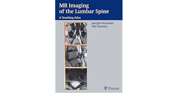
There is also a brief description of surgical technique and strategy. Additionally, the case studies provide great detail about clinical presentation, treatment, operative findings and clinical course. The title of the atlas is somewhat misleading, as this is an atlas of degenerative lumbar spine disease, and not an all-encompassing MRI atlas of lumbar spine pathology.
Although not an especially sexy topic, degenerative diseases of the lumbar spine afflict millions of patients. To some degree, practicing radiologists and those in training need to be proficient in the appropriate diagnosis of degenerative lumbar spine pathology. MR Imaging of the Lumbar Spine: A Teaching Atlas is a quick read that provides an extremely thorough and well illustrated review of degenerative lumbar spine disease.
Knake is a fourth year radiology resident at the University of Virginia in Charlottesville. He will be a musculoskeletal radiology fellow at the university in July To purchase this book, click here. If you are interested in reviewing books, let us know at residents auntminnie. The opinions expressed in this review are those of the author, and do not necessarily reflect the views of AuntMinnie.
If you like this content, please share it with a colleague! To read this and get access to all of the exclusive content on AuntMinnie.