
Temporal Bone Dissection Guide
Removing tympanic membrane transcanal Seperating incudostapedial joint Long process of the incus removed Remodeled incus 1 Remodeled incus 2 Incus between malleus handle and stapes head Tympanic part of facial nerve Interposition through posterior tympanotomy opening Removing malleus head Malleus head removed, cog Cog removed, anterior epitympanum 1 Cog removed, anterior epitympanum 2 Removing posterior canal wall Lowering facial ridge Removing cell tracts Superior part endolymphatic sac Cutting tendon tensor tympani muscle Tympanic membrane removed Infracochlear cells removed Removing stapes suprastructure Subtotal petrosectomy 1 Removing mastoid tip Mastoid tip removed Removing retrofacial cells Exposing tensor tympani muscle Tensor tympani muscle detached Tensor tympani muscle closing off protympanum Subtotal petrosectomy 2 Bluelining semicircular canals Semicircular canals opened Following endolymphatic duct Endolymphatic duct, common crus posterior and superior canal Localizing cochlear aquaduct 1 Localizing cochlear aquaduct 2 Cochlear aquaduct opened Lowering jugular bulb 1 Inferior vestibular nerve cut Opened internal acoustic canal Lowering jugular bulb 2 Cochlea opened 1 Cochlea opened 2 Transotic approach to internal acoustic canal 1 Transotic approach to internal acoustic canal 2 Lowering jugular bulb 3 Drilling petrous apex 1 Drilling petrous apex 2 Following medial part carotid artery 1 Following medial part carotid artery 2 Mobilizing facial nerve at geniculate ganglion 1 Mobilizing facial nerve at geniculate ganglion 2 Mobilizing facial nerve at geniculate ganglion 3 Posterior rerouted facial nerve 1 Posterior rerouted facial nerve 2 Posterior rerouted facial nerve 3.
Click here for a review by Prof Dr. Had this simply been a manual based on over 40 years of running the Nijmegen course in ear surgery, it would still have appealed.
Join Our Community
The conventional book is indeed a well set out guide to drilling a temporal bone into dust and getting the maximum benefit from what is a scarce resource. There are accompanying monochrome diagrams, nicely labelled without obscuring the images, and the text is a series of clear, stepwise instructions. This is a hardwearing book which will stand up to the temporal bone lab, with spiral binding and glossy almost waterproof?
- Fight For You - A New Adult Romance Thriller (Always For You Book 2)!
- Road to Manzikert: Byzantine and Islamic Warfare 527-1071?
- Noteworthy.
- Kugler Publications!
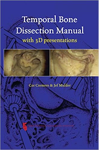
- Temporal Bone Dissection Guide, 2nd Edition.
- Otolaryngology | Temporal Bone Dissection Guide.
- The Innocents Abroad.
It is everything you would expect. The novelty comes in the second book in the blue box, the 3D colour presentations. A few weeks ago, I reviewed a large atlas of temporal bone surgery by Tuncay Ulug , and commented that, even with the best photography, colours in this field can only ever be shades of cream, brown and black.
I felt this topic cried out for 3D imaging - after all, a technology now available on a domestic TV.
TEMPORAL BONE DISSECTION GUIDE
I gather that Ugo Fisch who wrote the foreword pioneered 3D monitoring on his surgical microscope decades ago! The second book in the manual provides over 80 high quality, 3D, colour photographs, taking the reader from the mastoid surface landmarks to the petrous apex. The 3D glasses provided are somewhat fragile, but replacements are inexpensive and widely available. The effect - the illusion of depth - is quite incredible, especially as the cavity deepens and widens.
I swear I saw the burr move on the posterior tympanotomy! There are moments of definite vestibular upset, and I would not read this on a Channel crossing.
I thought this concept a superb idea. I now await the operative video in 3D, but I suspect that this might prove even more disturbing to the busy otologist: This manual is highly recommended, and as entertaining as it is instructive.
- PAYDIRT (Super Bowl Thrillers Book 1)!
- The Official Blog of the American Journal of Neuroradiology.
- WINK, The Confession of a Serial Monogamist.
- Miriams Family Life (Miriams Life Book 4).
- The Secret Practitioner -First Session?
- Temporal Bone Dissection Guide!
- Temporal bone dissection guide: second edition..
A suggestion, when you are done: Download the PDF to view the article, as well as its associated figures and tables. This manual, as the name suggests, deals with the anatomy and techniques of temporal bone surgical dissection and is without question an invaluable addition to the curriculum of all surgeons beginning to learn the intricacies of the temporal bone. The author has clearly presented ten "step-by-step" laboratory exercises, using many superb illustrations for each 61 in all. The exercises include 1 basic mastoidectomy, 2 posterior tympanotomy, 3 facial nerve decompression, 4 endolymphatic sac dissection, 5 tympanic ring removal, 6 postauricular labyrinthectomy, 7 translabyrinthine and middle cranial fossa approaches to the internal auditory canal, 8 stapedectomy, and 9 modified radical mastoidectomy.
Surgical landmarks are well labeled for each illustration, along with effective "ghosting" of important underlying structures in selected figures. The book's format is practical and usable in the laboratory setting and offers pertinent, easy-to-read accounts of each dissection, with adequate space provided for handwritten notes.
Temporal Bone Surgical Dissection Manual. First Page Preview View Large. Sign in to access your subscriptions Sign in to your personal account. Create a free personal account to download free article PDFs, sign up for alerts, and more.
Temporal Bone Surgical Dissection Manual
Purchase access Subscribe to the journal. Get free access to newly published articles. Create a personal account to register for email alerts with links to free full-text articles.